G2107
Sclerotinia Diseases of Sunflower in Nebraska
Sclerotinia is often considered the most serious pathogen of sunflowers. This NebGuide discusses signs, symptoms, and management of this disease.
Robert M. Harveson, Extension Plant Pathologist
- Introduction
- Diseases, Signs, and Symptoms
- Basal Stalk Rot and Wilt
- Mid-Stalk Rot
- Head Rot
- Disease Cycle
- Management
|

Introduction
Sclerotinia sclerotiorum is a common and widespread soilborne fungal pathogen. It was first described in 1837, and recognized as a sunflower pathogen in 1861. Within temperate regions of the world, it is often considered the most serious pathogen of sunflower because it is capable of infecting multiple organs (roots, stems, buds, and heads); persists for many years in soils; and has a large host range consisting of more than 360 plant species belonging to 225 genera in 64 families. Sclerotinia minor is another species reported from South America, Australia, and California causing root rot and wilt on sunflower, but is much less commonly found than S. sclerotiorum. The two pathogens can be differentiated on the basis of the size of the sclerotia (overwintering structures). S. minor has uniformly round sclerotia measuring 0.5 to 2 mm (1/12 to 1/40 of an inch) while those of S. sclerotiorum produce larger and irregular sclerotia, some measuring 1 to 5 cm (0.5 to 2 inches). Sclerotia produced by S. sclerotiorum in heads are very similar in size and shape to sunflower seeds (Figure 1).
Diseases, Signs, and Symptoms
Significant yield reductions have been documented in all regions of the world. However, the impact of the pathogen on yield is determined by the type of disease and symptoms observed in fields. S. sclerotiorum can induce three distinctly different diseases on sunflower: 1) basal stalk rot and wilt; 2) mid-stalk rot; and 3) head rot, while S. minor only incites the basal stalk rot and wilt disease.
Basal Stalk Rot and Wilt
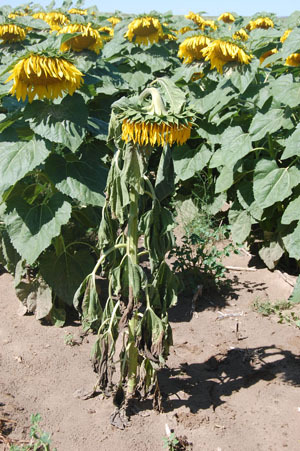
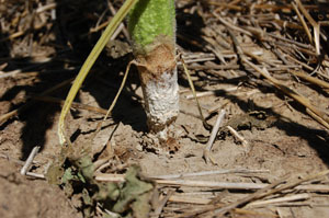
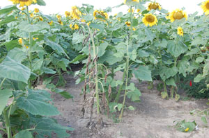
Each of the three Sclerotinia diseases will produce distinct symptoms. Basal stalk rot can occur anytime after the seedling stage but is usually more prevalent during flowering (Figures 2 and 3). Basal stalk rot is triggered through root infection from the pathogen residing in the soil. The first symptom seen aboveground is a sudden wilting of the entire plant without any foliar lesions or spots (Figure 4). A light-brown, water-soaked lesion will be evident at the soil line (Figure 5). If moisture conditions remain conducive, a white, cottony mycelial growth will be observed on lesions at the soil line (Figure 6), and may rapidly girdle and extend up the stalk 4-6 inches above the ground (Figure 7). With time the fungus grows internally, digesting the pith, and causing shredding and bleaching of infected stems (Figure 8). The most diagnostic trait of the stalk rot disease is the hollowed-out, rotten stalks filled with black sclerotia (Figure 9). Late infections or those causing less severe invasion may exhibit the white mycelium at the soil line without wilting. However, these types of infections still weaken the plant, making them more susceptible to lodging (Figure 10).
|
 |
|||
| Figure 3. Basal stalk/root rot infection after flowering, but before maturity | ||||
 |
 |
|||
| Figure 4. Basal stalk/root rot early infection showing permanent wilting symptoms | Figure 5. Water-soaked lesions at base of stalk | |||
 |
 |
|||
| Figure 6. White mycelium (mold) at base of infected sunflower plant | Figure 7. White mycelium extending up the stalk above the soil line under high humidity (Courtesy of Tom Gulya, USDA-ARS, Fargo, North Dakota) | |||
|
 |
|||
| Figure 9. Diagnostic signs and symptoms of Sclerotinia wilt consisting of numerous black sclerotia forming in hollowed-out pith of stalk | ||||
|
||||


Mid-Stalk Rot
Mid-stalk rot is caused only by S. sclerotiorum and generally occurs between the late vegetative stage and maturity via ascospores discharged from mushroom-like apothecia (Figure 11). It is not as commonly observed in Nebraska as the basal stalk rot and wilt disease phase. Although infection may start in leaf axils, it more commonly begins as a leaf infection (Figure 12) before progressing down the petioles to stalks. Ascospore infections produce lesions on stems (Figure 13), similar to those at the soil line from basal stalk rot and wilt. Mid-stalk infections may not cause wilting or death of plants, but will often result in breakage of stems at the point of infection (Figure 14), resulting in complete yield loss in affected plants.
 |
 |
|||
| Figure 11. Cup-shaped apothecia that produce infective ascospores, formed in field | Figure 12. Foliar infection of S. sclerotiorum from ascospores. Note the senescent flower petal on leaf that served as a nutrient source for initiating infection by ascospores (Courtesy of Sam Markell, North Dakota State University) | |||
 |
|
|||
| Figure 13. Mid-stalk rot infection (arrow) from airborne ascospores (Courtesy of Tom Gulya, USDA-ARS, Fargo, North Dakota) |

Head Rot
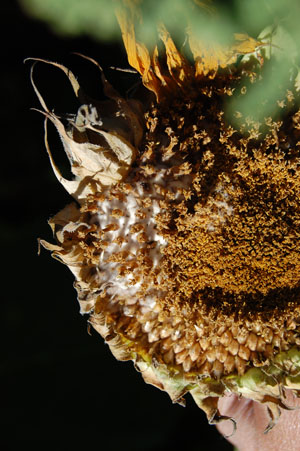
Head rot usually occurs at the end of flowering or later, but also may be found before flowering (referred to as bud rot). Initial symptoms may consist of dark, water-soaked spots on the back side of the heads (similar to the beginning of Rhizopus head rot), or white mycelial growth covering portions of the developing seeds (Figure 15). As the disease progresses, the fungus rots the interior of the head and large numbers of irregularly shaped sclerotia fill the head. Sclerotia are often perforated by developing seeds, giving the remaining head tissue a net-like appearance (Figure 16). The disease eventually results in compete disintegration of heads, leaving nothing but frayed vascular elements resembling a broom head (Figure 17).
 |
|
|||
| Figure 15. White mycelial growth (mold) characteristic of Sclerotinia head rot | ||||
 |
 |
|||
| Figure 17. Completely rotted head consisting only of vascular elements resembling a broom head (Courtesy of Tom Gulya, USDA-ARS, Fargo, North Dakota) | Figure 18. Germinating sclerotia in culture. Note numerous cup-shaped apothecia arising from each sclerotium. |

Disease Cycle
Both Sclerotinia species (S. sclerotiorum and S. minor) overwinter in the soil as resistant, hard-walled sclerotia. Diseases incited in sunflowers by Sclerotinia have two different disease patterns, both of which begin with the sclerotia. One involves the mycelial germination of the sclerotia, which results in root infections and the development of the basal stalk rot and wilt disease, while the other pattern involves the production of apothecia and ascospores from sclerotia, which then induce the mid-stalk and head rot diseases.
Sclerotia of both species are stimulated to germinate and produce mycelium by root exudates. Optimal temperatures for mycelial growth are 75-78°F, but infection cannot occur unless the pathogen comes into contact with plant roots. The pathogen then penetrates into roots and invades both down and up within stems, decomposing all tissues it contacts as it grows. Sclerotia are formed from white, cottony hyphae on root surfaces, basal portions of the stem, and inside the hollow pith of affected stem tissues where they return to the soil after plants decompose. Sclerotia can survive at least five years under cool and dry soil conditions. Soil temperatures above 80°F with high moisture levels are not favorable for survival or maintaining viability of sclerotia.
Sclerotia of S. sclerotiorum that remain close to the soil surface can germinate and form the mushroom-like apothecia that produce and release ascospores. One to several mushroom-like apothecia can be formed per sclerotium (Figure 18). The ascospores are short-lived once released but can be disseminated long distances (one-half mile). S. minor does not produce apothecia or ascospores. Apothecial production and ascospore release occur at a temperature range of 40-90°F, but spore germination and infection of sunflowers is optimal at 75-78°F. Infection by ascospores requires both free water and an energy source, usually supplied by wounded or senescing tissues. Ascospores can remain viable for up to 45 days at temperatures of 65-75°F and low humidity, and can remain viable for several months at 40°F.
Apothecial formation is favored by high soil moisture resulting from high rainfall levels. Sclerotia buried deeper than 0.75 to 2 inches cannot produce apothecia that will reach the soil surface, and the sclerotia closest to the surface have the highest frequency of apothecia production.
Management
Sclerotinia diseases are traditionally very difficult to control due to the large host range and longevity of sclerotia in soil. The most effective methods of control integrate cultural practices, fungicide use, and genetic resistance.
- Shallow cultivation of reduced tillage may help reduce viability of sclerotia exposed to cold winters in North America.
- Deep cultivation will bury sclerotia and increase chances of survival and root infections, but also will reduce sclerotial numbers near the soil surface and reduce chances of ascospore infection of heads and midstems.
- Rotations of three to five years with nonhost monocots will reduce numbers of sclerotia in soil, thereby reducing chances of root and wilt infections.
- Plant resistant cultivars where possible.
- Chemical (fungicide) control of foliar infections from ascospores has been demonstrated to be successful and economical if properly timed.
This publication has been peer reviewed.
Visit the University of Nebraska–Lincoln Extension Publications website for more publications.
Index: Plant Diseases
Sunflower
Issued September 2011