G1957
Bacterial Brown Spot of Dry Beans in Nebraska
Bacterial brown spot and its management are covered in this NebGuide.
Robert M. Harveson, Extension Plant Pathologist
Introduction
Bacterial brown spot is a more recently discovered disease of dry beans in Nebraska than either halo blight or common bacterial blight. It was first seen in western Nebraska dry bean fields on a limited basis throughout the late 1960s. Epidemics still occur sporadically, but the pathogen’s presence has increased in incidence and damage along with halo blight during the past 20 years in the Central High Plains.
Brown spot-tolerant bean cultivars were first identified and reported by Schuster and Coyne in 1969 from field and greenhouse tests in Nebraska using the dry bean lines US 1141 and GN Nebraska #1 selection 27, and the snap bean cultivar Tempo. When the disease occurs today, however, it can be very damaging due to the lack of resistance in modern commercial cultivars.
Symptoms and Signs
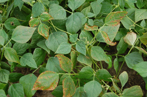
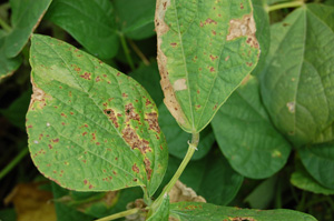
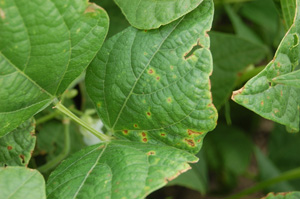
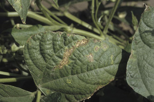
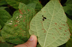
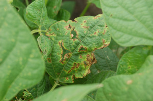
Lesion size can vary, but generally lesions are small, circular, and brown (Figure 1), often surrounded by a yellow zone (Figure 2). As the disease progresses, lesions begin combining to form linear, necrotic streaks bound by leaf veins (Figure 3). If water soaking occurs, it appears as small circular spots on the underside of leaves (Figure 4). Old lesion centers fall out, leaving tattered strips or “shot holes” on affected leaves (Figure 5) and evidence of water soaking may be visible in the edge of tissue next to the shot holes. Stem and petiole lesions are occasionally found when the pathogen becomes systemic.
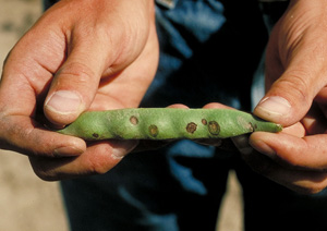
Pod lesions are circular and water-soaked initially, but later turn brown and become necrotic (Figure 6). If young pods or those in the flat stage become infected, they may be bent or twisted with ring-spots or water-soaked brown lesions.
Like the halo blight pathogen, a cream to silver-colored bacterial ooze may emerge from stems, pods or leaves after infection under humid climatic conditions. Seeds may also shrivel or discolor, or become infected if lesions penetrate into pod walls.
 |
 |
|
| Figure 1. Small, brown, necrotic lesions characteristic of early bacterial brown spot infection. | Figure 2. Bacterial brown spot lesions surrounded by yellow zones. | |
 |
 |
|
| Figure 3. Bacterial brown spot lesions coalescing to form linear necrotic lesions between veins. | Figure 4. Water-soaking symptoms of bacterial brown spot on underside of leaves (leaf on right). | |
 |
 |
|
| Figure 5. Areas of leaf where dead centers of old bacterial brown spot lesions have fallen out leaving tattered holes or strips. | Figure 6. Dried, necrotic bacterial brown spot lesions on pods. Credit: H.F. Schwartz. |
Pathogen and Disease Cycle
Pseudomonas syringae pv. Syringae (Pss) is a strictly aerobic or oxygen-requiring bacterium capable of movement. Like Psp (the halo blight pathogen), it produces diffusible, fluorescent pigments on iron-deficient media, and cream to white colonies on standard media (Figure 7). Pathogenic isolates may produce a bacteriocin (compound toxic to other related bacteria) in the host plant.
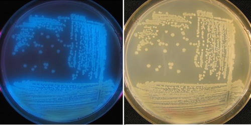 |
| Figure 7. Fluorescent growth of brown spot pathogen on iron-deficient media under a black light (left); white to cream colored growth on standard media (right). Credit: H.F. Schwartz. |
The pathogen Psuedomonas syringae causes disease on many different plants, but only certain forms of the pathogen cause brown spot. Pss has been demonstrated to be extremely variable among leguminous hosts. For example, isolates from cowpeas (black-eyed peas) were shown to be weaker parasites than those from the original New Jersey bean specimens. The isolates originally found in Nebraska dry bean production areas were shown to be less infectious than snap bean isolates from Wisconsin. Isolates from canning peas in Wisconsin were able to produce brown-spot symptoms on beans and vice versa. Furthermore, its pathogenic host range is very large, including Phaseolus vulgaris (common bean), P. lunatus (lima bean), Pueraria lobata (kudzu), Glycine max (soybean), Pisum sativa (pea), Vicia faba (fava bean), Vigna sesquipedalis (yardlong bean), Vigna unguiculata (cowpea) and V. sinensis (mung bean).
Bacterial brown spot, like common bacterial blight, is a warm weather-oriented disease. The pathogen potentially causes the most damage when temperatures range from 80°F to 85°F (27°C to 30°C), as opposed to the cool weather favoring disease development of halo blight (68°F to 72°F; 17°C to 20°C).
|
Conditions for brown spot development in Nebraska often occur during mid-vegetative (Figure 8) to early-flowering periods of plant growth. The pathogen may only need cool nights to thrive.
Like other dry bean bacterial pathogens, the brown-spot pathogen survives in bean residue or seeds from the previous year, although seed infections are not thought to be important in the epidemiology of the disease except to introduce the pathogen into new areas. The pathogen has additionally been shown to have a resident phase on both host and nonhost plants. Thus, the pathogen is also capable of growing and surviving as an epiphyte (an organism that can survive on the external leaves of another plant) on the surface of both weed and legume crop hosts. Wet weather, hail, violent rain and windstorms help the pathogen to spread among and between fields.
Management
Chemical Methods
Chemical control results vary depending on pathogen, weather and disease pressure. Increased yields have been realized for halo blight and brown spot infections through use of copper-based applications 40 days after emergence (mid-vegetative or early flowering periods) and then repeated every seven to 10 days for a total of three applications.
Cultural Methods
- Do not save seed from previously blighted fields for re-use.
- Plant certified seed of disease-resistant cultivars where possible.
- Plant seed treated with streptomycin to help reduce contamination of the seed coat and establish a vigorous early-season stand.
- Rotate beans with other crops for two to three years.
- Incorporate infected bean residues after harvest.
- Eliminate bean volunteers during growing season.
- Stay out of bean fields when wet. Moving through blighted fields when foliage is wet can spread pathogen to other plants.
- Do not spread old bean straw from infested crops on new fields to be used for bean production.
- Avoid re-using irrigation water.
- Do not plant beans close to recently blighted fields.
This publication has been peer reviewed.
Visit the University of Nebraska–Lincoln Extension Publications Web site for more publications.
Index: Plant Diseases
Dry Beans
Issued July 2009
