G1677
Rhizopus Head Rot of Sunflower in Nebraska
Sunflower crops grown in Nebraska are susceptible to diseases caused by bacteria, viruses, and nematodes, but the most economically damaging is head rot, caused by the fungal pathogen Rhizopus.
Robert M. Harveson, Extension Plant Pathologist
|
Introduction
Sunflower production in Nebraska and other areas of the central High Plains (Colorado and Wyoming) has been on the increase in the last decade due, in part, to the establishment of oil and confectionary seed industries in local markets. The crop is well-adapted to this region and can be successfully cultivated in both dryland and irrigated areas.
Reported diseases of sunflowers include those caused by fungi, bacteria, viruses, and nematodes. However, most common diseases affecting sunflowers in this region are caused by fungi and are generally more severe in irrigated production.
The Pathogen and Disease History
Rhizopus is a common fungus that occurs naturally in soils and as airborne spores. Head rot disease is caused by several species of this genus, including R. stolonifer, R. arrhizus, R. oryzae, and R. microsporus. These species may occur singly or in a complex. This genus is well-known for causing soft rots of fruits, vegetables, and root crops, especially in postharvest storage situations.
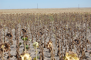
Rhizopus head rot is one of the few diseases caused by this group of fungi that occurs in field crops during the cropping season. Historically it has been of minor importance in the United States and Canada; however, in a recent survey of sunflower diseases in California, Rhizopus head rot was found to be the most severe and commonly identified disease. It also has been documented to cause severe damage annually to sunflower grown in Israel, and has been shown to be a very serious disease in the High Plains under the right environmental conditions (see Disease Cycle and Damage), causing up to 100 percent losses in severely affected fields (Figure 1).
Symptoms
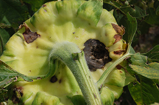
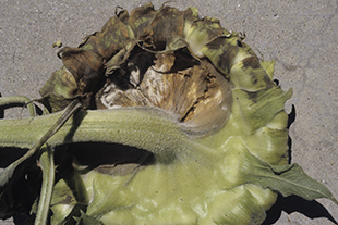
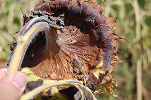
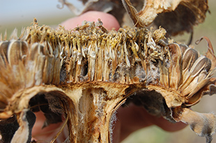
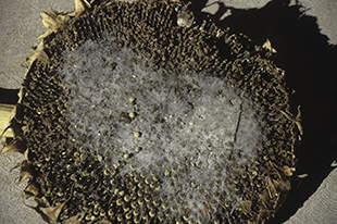
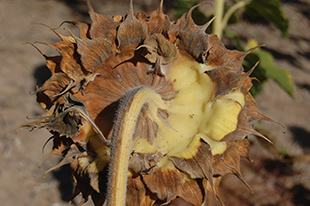
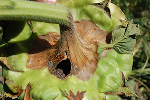
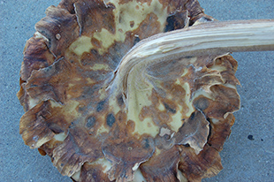
The disease first appears as dark spots on the back of ripening heads (Figure 2), followed by a watery, soft rot that later turns dark brown (Figure 3). As disease progresses, heads dry prematurely, shrivel, and tissues appear to shred (Figure 4). The fungus can be observed inside shredded heads as coarse, threadlike mycelial strands (Figure 5) that are later followed by the appearance of fungal reproductive structures (fruiting structures) called sporangia that look like small black dots about the size of pinheads. The disease also can be recognized on the flower side of the head by the appearance of a grayish, fuzzy substance (Figure 6) covered with sporangia. These dark fruiting bodies are sacks filled with spores that are easily broken, thereby spreading the spores to neighboring plants by wind. Symptoms can often seem to appear simultaneously on opposite sides of the head. The fungus also causes a systemic effect in the plant on the side where the head became infected (Figure 7). If the peduncle becomes infected, it can allow the head to fall completely off, increasing the potential for severe yield loss.
 |
 |
|
Figure 2. Wounds on the back of the head providing an entry point for infection (necrotic areas) by the pathogen. |
Figure 3. Initial watery, soft rot, followed by dried, brown tissues. |
|
 |
 |
|
Figure 4. The disease progresses, resulting in dried tissues that begin to shred. |
Figure 5. Severely infected head showing grayish hyphae growing through the head. |
|
 |
 |
|
Figure 6. The pathogen growing through the head and emerging on the flower side of the head after initial infection occurred on the back. |
Figure 7. One-sided rot moving down the stem from the affected side of the head. |
Disease Cycle and Damage
Even though Rhizopus spp. spores are found everywhere in the environment, mechanical injury on heads is a prerequisite for infection and disease development (Figure 8). Afterwards, the disease becomes more severe in warm, humid environments, especially under irrigation. Thus, as spores are carried to sunflower plants through air currents, and under conditions of high humidity, infection is initiated through wounds created by hail (Figure 9), birds, or insects.
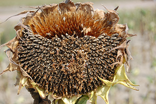
Damage and economic losses depend on the creation of wounds, the environmental conditions present, and the time of season that wounding and infection occur. Research has demonstrated that head rot rarely occurs before flowering, so it appears that mature tissues are required to support the growth of Rhizopus spp. Yields are negatively affected because seeds in infected heads fail to fill properly and have reduced weights. This can obviously affect profits for confectionary seed growers since payment is based on seed size. Yield also can be affected even if infection occurs late in the season due to the loss of seeds that have fallen to the ground from affected heads (Figure 10). Oil seed growers also may be adversely affected by head rot due to bitter or poor quality oils obtained from infected plants.
 |
 |
|||
Figure 8. Rot progressing on the head from the large wound (hole). |
Figure 9. Severe hail damage that initiated the disease on the head. Note necrosis affiliated with the circular wounds. |
|||
|
||||
Management
- Avoid mechanical damage after flowering.
- Control head moth infestation before or at flowering.
- Remove wild sunflowers that may serve as reservoirs for insects and pathogens before they produce seed.
- Control bird feeding:
- Select varieties with head types that turn down after flowering.
- Avoid planting sunflowers near water that consistently harbors many birds.
This publication has been peer reviewed.
Visit the University of Nebraska–Lincoln Extension Publications website for more publications.
Index: Plant Diseases
Field Crops
2007, Revised October 2013